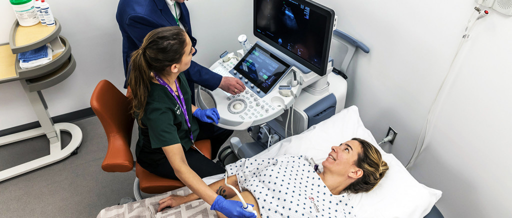
What is Routine Sonography ?
Routine sonography is a diagnostic imaging technique that uses high-frequency sound waves to produce images of the body’s internal organs. It is commonly used to assess the fetus’s health during pregnancy, and can also be used to detect abnormalities and diagnose diseases in adults. Sonography is a non-invasive procedure and does not involve exposure to radiation. Dr. Prajakta Patil provides routine sonography services in Dombivli.
The images produced can help physicians diagnose a variety of medical conditions. Sonography is non-invasive, meaning it doesn’t involve the use of needles or other instruments that penetrate the skin. This makes it a relatively safe and comfortable procedure for most patients. Additionally, sonography produces clear images that can be easily interpreted by doctors. This makes it an effective tool for diagnosing a wide range of medical conditions. We provide various sonography services in Dombivli.
Abdomen & Pelvis: Abdomen sonography is a common but important tool for detecting various diseases of the abdominal organs.
Kidney and Urinary bladder: Kidney and urinary bladder sonography is a type of imaging that uses sound waves to produce pictures of organs in the body.
Chest: Chest sonography is an important diagnostic tool for evaluating patients with chest pain.
Pelvis: Pelvic sonography is a procedure that uses sound waves to image the internal organs in the pelvis.
Full Bladder: A full bladder sonography is a type of diagnostic imaging that uses sound waves and pictures to look inside the bladder and determine if it is full. This test can help identify problems, such as urinary tract infections, that may be causing pain or difficulty urinating.
